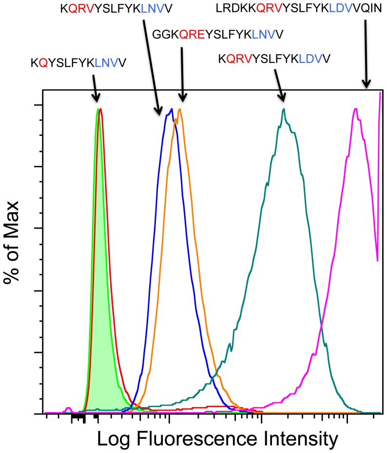
Figure 9. The QRV/QKE motif rescues binding of peptides with a mutated canonical α4β7 binding site.
The graph shows the histogram peaks generated by binding of short biotinylated peptides to α4β7+ RPMI 8866 cells. The binding activity of the short peptide with a mutated α4β7 binding site (LNV instead of LDV) was restored with the addition of QRV (blue) or QKE (orange) to the N-terminus of the peptide. In the diverse set of peptides tested, maximal binding was achieved with the inclusion of both the QRV and LDV motifs (green and magenta). Green histogram is NeutrAvidin control, LFI = Log Fluorescence Intensity.

