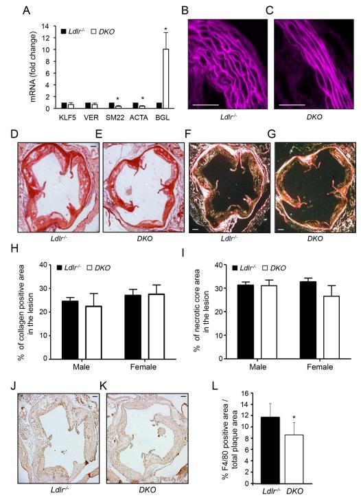
Figure 2. Atherosclerotic plaque structure and composition in Ldlr−/− and DKO mice.
A) qRT-PCR analysis of KLF5, Versican (VER), SM22, ACTA and BGL expression in aortic tissues isolated from Ldlr−/− and Ldlr−/−-miR-143/145−/− (DKO) mice. Data are expressed as mean ± S.D. * p<0.05 vs. Ldlr−/−. B and C) Representative histological images of the media structure of the thoracic aorta isolated from. Ldlr−/− and DKO mice. D-G) Representative histological analysis of cross-sections of the aortic sinus from Ldlr−/− and DKO mice stained with sirius red (D and E) and following acquisition with polarized light (F and G). H and I) Quantification of the collagen content (H) and necrotic areas (I) in the aortic sinus. Data are presented as the percentage of atherosclerotic lesion covered by collagen or the necrotic core, and are expressed as mean ± S.D. (n=8 for each group). J and K) Representative F4/80 stained aortic sinus sections from Ldlr−/− and DKO mice. L) Quantification of the F4/80 positive areas within the atherosclerotic plaques. Data are expressed as mean± S.D (n=8 for each group). Scale bar indicates 100μm.

