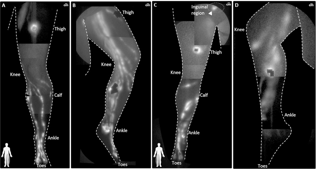
Figure 3.
NIRF lymphatic imaging montages of the (A) anterior aspect and (B) medial aspect of the lymphatics in the left leg of NC3 (BMI 30.2) and (C) the anterior aspect and (D) medial aspect of the lymphatics in the right leg of DD3 (BMI 30.9). The leg lymphatics of DD3 are dilated in the anterior shin and a painful, fluorescent, fibrotic mass (arrowhead) was found in the inguinal region. The lymphatics in the medial calf are dilated and correspond with fibrotic and nodular tissues thought to be lymphatic vessels. Injection sites are covered by round bandages and/or black vinyl tape. See Supplemental Videos 3 and 4, for a comparison of functional lymphatic drainage in participants NC3 and DD3.

