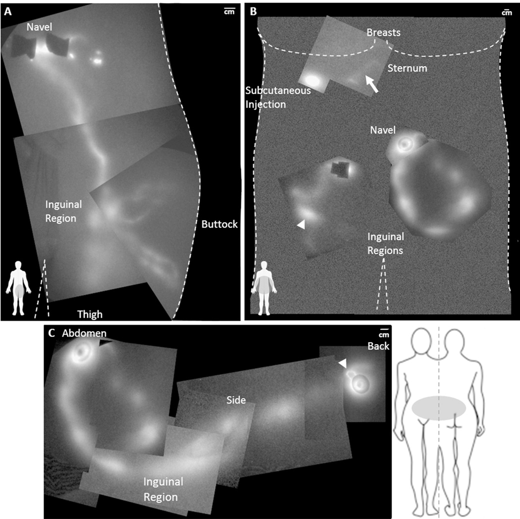
Figure 5.
(A) NIRF lymphatic imaging montage of the lymphatic drainage to the inguinal region from intradermal injections near the navel, above and below the buttock, and from the leg in NC1 (BMI 25.0). (B) Montage of the lymphatic drainage of the abdomen of DD3 (BMI 30.9). The arrow identifies the location of a dim lymphatic vessel draining from the subcutaneous injection below the right breast towards the sternum. The arrowhead identifies the location of a fluorescent, painful fibrotic mass palpated in the right inguinal region and corresponds to the arrowhead in Figure 2C. (C) NIRF lymphatic imaging montage illustrating the lymphatic drainage pattern from the intradermal injections on the abdomen and lower back to the inguinal region of DD3. The arrowhead identifies abnormal lymphatic capillary structures near the injection site on the back. Note the segmented appearance of the lymphatics. Injection sites are covered by round bandages and/or black vinyl tape.

