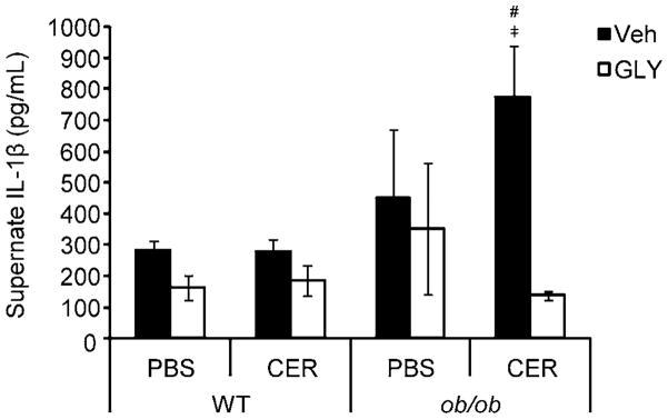
Figure 5. LPS-stimulated IL-1β production by ex vivo peritoneal cells from WT and ob/ob mice following cerulein-induced AP with or without glyburide treatment.

AP was induced in lean WT and obese ob/ob mice via eight (8) hourly IP injection of cerulein (CER; 100 μg/kg or PBS (v/v) control) following glyburide (GLY; 500 mg/kg) or vehicle (Veh; v/v) treatment. Following sacrifice, peritoneal cells were collected via lavage, washed, plated, stimulated with LPS (1 μg/mL) and incubated overnight. Supernatents were collected for analysis of IL-1β concentration by ELISA. Results are expressed as mean ± s.e.m. n = 3–14 mice per group. #P < 0.05 vs. respective WT group. ╪P < 0.05 vs. respective glyburide treated group.
