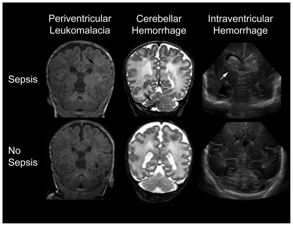
Figure 1.
Representative T1-weighted image of periventricular leukomalacia (left), T2-weighted MR image of cerebellar hemorrhage (middle), and coronal ultrasound images of intraventricular hemorrhage (right). Images from infants exposed to sepsis (top) have arrows indicating the relevant pathologies. The bottom images are from a very preterm infant without any infectious exposure.

