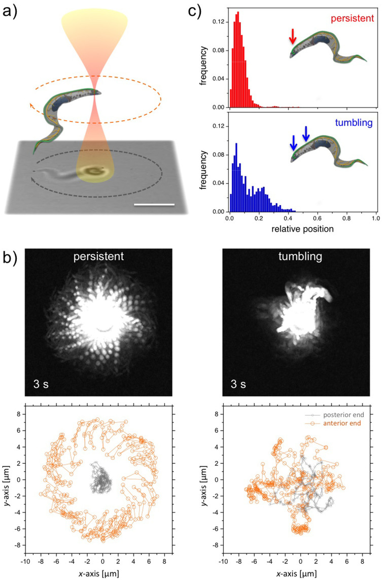
Figure 1.
Optical trapping of trypanosomes: (a) Schematic representation of an optical tweezers setup for trapping motile trypanosomes. Within the optical trap the motility is limited, whereas the mobility is unaffected. (b) Overlay of images (3 s, frame rate 100 Hz) from persistent and tumbling walkers in the optical trap. Trajectories of the posterior and anterior end of persistent and tumbling trypanosomes are displayed in the lower part. (c) Histograms of tapping loci of persistent walker and tumbling cells versus the position from the posterior end relative to their contour length L. The main trapping positions indicated by arrows are at the posterior end close to the flagellar pocket.

