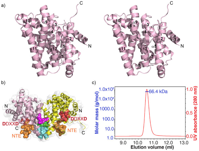
Figure 2. Structure of BjKS.
(a) Stereoview of (apo-1) BjKS (PDB ID code 4KT9) showing highly α-helical structure. (b) Dimer interface in B. japonicum ent-kaurene synthase. Hydrophobic residues (M235, Y232 and F240, cyan spheres) form a hydrophobic core; E244 side-chains interacts with the N-terminus of the helix starting at Y155 in the other subunit (green spheres); salt bridges exist between the E157(A) and R250(B) side-chains, and between E157(B) and R250(A) (purple spheres). The two catalytic motifs (DDXXD, red and NTE, orange) are also shown. (c) SEC-MALS results.

