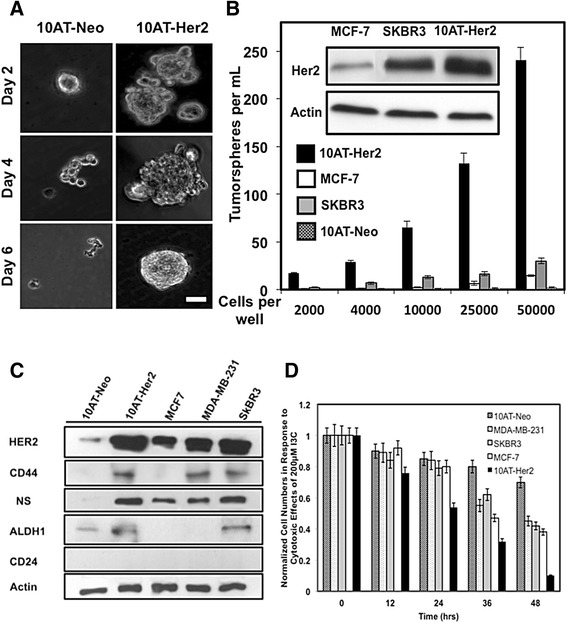
Figure 2.

Tumorsphere formation efficiency in cell suspension cultures, expression of stem/progenitor cell marker proteins and proliferation of breast cancer cells. (A) 10AT-Her2 and 10AT-Neo cells were plated at a density of 4,000 cells per well in serum-free non-adherent suspension cultures as described in the Methods section. At the indicated days in culture, tumorsphere formation was assessed visually by phase microscopy. Scale bar represents 50 μm. (B) 10AT-Her2 and 10AT-Neo cells as well as the SKBR3 and MCF-7 human breast cancer cell lines were incubated at the indicated cell densities. Tumorsphere formation efficiency was quantified after 6 days in culture under non-adherent conditions. The presented values are an average of three independent experiments. The gel inserts are Western blots showing relative levels of HER2 protein expression and actin controls from electrophoretically fractionated total cell extracts of MCF-7, SKBR3 and 10AT-Her2 cells. (C) Cultured 10AT-Neo, 10AT-Her2, MCF-7, MDA-MB-231 and SKBR3 cells were harvested, total cell extracts electrophoretically fractionated and the levels of expressed HER2, CD44, CD24, ALDH-1, nucleostemin (NS) and actin protein determined by Western blots. (D) To examine the effects of I3C on cell proliferation, 10AT-Neo, 10AT-Her2, SKBR3, MCF-7 and MDA-MB-231 cells cultured in a 24-well plate were treated with 200 μM I3C for the indicated times and the cell number quantified as described in the Methods section. ALDH-1, aldehyde dehydrogenase-1; HER2, human epidermal growth factor receptor-2; NS, nucleostemin.
