Figure 6.

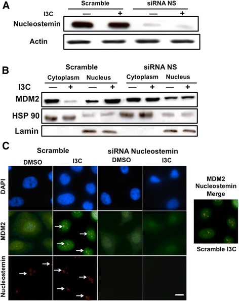
Nucleostemin-dependent I3C stimulation of MDM2 nuclear compartmentalization and localization into nucleolus foci. 10AT-Her2 cells were transfected with either control scramble siRNA or nucleostemin siRNA, and then treated with or without 200 μM I3C for 48 hours. (A) The level of nucleostemin protein was determined by Western blot analysis. (B) Cell extracts were biochemically separated into nuclear enriched and cytoplasmic fractions, electrophoretically fractionated, and Western blots probed with antibodies specific for MDM2, the cytoplasmic marker HSP90 and the nuclear marker lamin. (C) The subcellular localization of MDM2 and nucleostemin was determined by indirect immunofluorescence microscopy. DAPI staining was used to visualize DNA stained nuclei. Scale bar represents 4 μm. DAPI, 4',6-diamidino-2-phenylindole; DMSO, dimethyl sulfoxide; I3C, indole-3-carbinol; MDM2, murine double mutant 2; NS, nucleostemin; siRNA, small interfering RNA.
