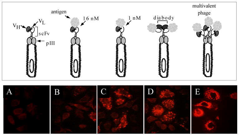
Figure 1. Phage antibodies are endocytosed into ErbB2 expressing cells.
Top panel shows different phage antibody constructs studied. Bottom panel shows immunofluorescent microscopy, staining for phage major coat protein pVIII. Panels A–D phage are displayed in a phagemid vector where there is a single scFv/phage. Panel E phage are displayed in a phage vector with 3–5 copies of scFv/phage. A. Control phage antibody (binds BoNT). B. C6.5 anti-ErbB2 scFv. C. Higher affinity ML3-9 anti-ErbB2 scFv. D. Dimeric C6.5 diabody. E. C6.5 scFv displayed multivalently in true phage vector.

