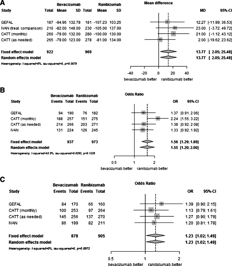
Fig. 3.
Comparison of bevacizumab with ranibizumab for anatomical results. Panel a, Change in retinal thickness at fovea at 1 year (microns); Panel b, Presence of intraretinal or subretinal fluid on optical coherence tomography at 1 year; Panel c, Presence of dye leakage on angiogram at 1 year. CI, confidence interval; MD, mean difference; OR, Odds ratio; SD, standard deviation

