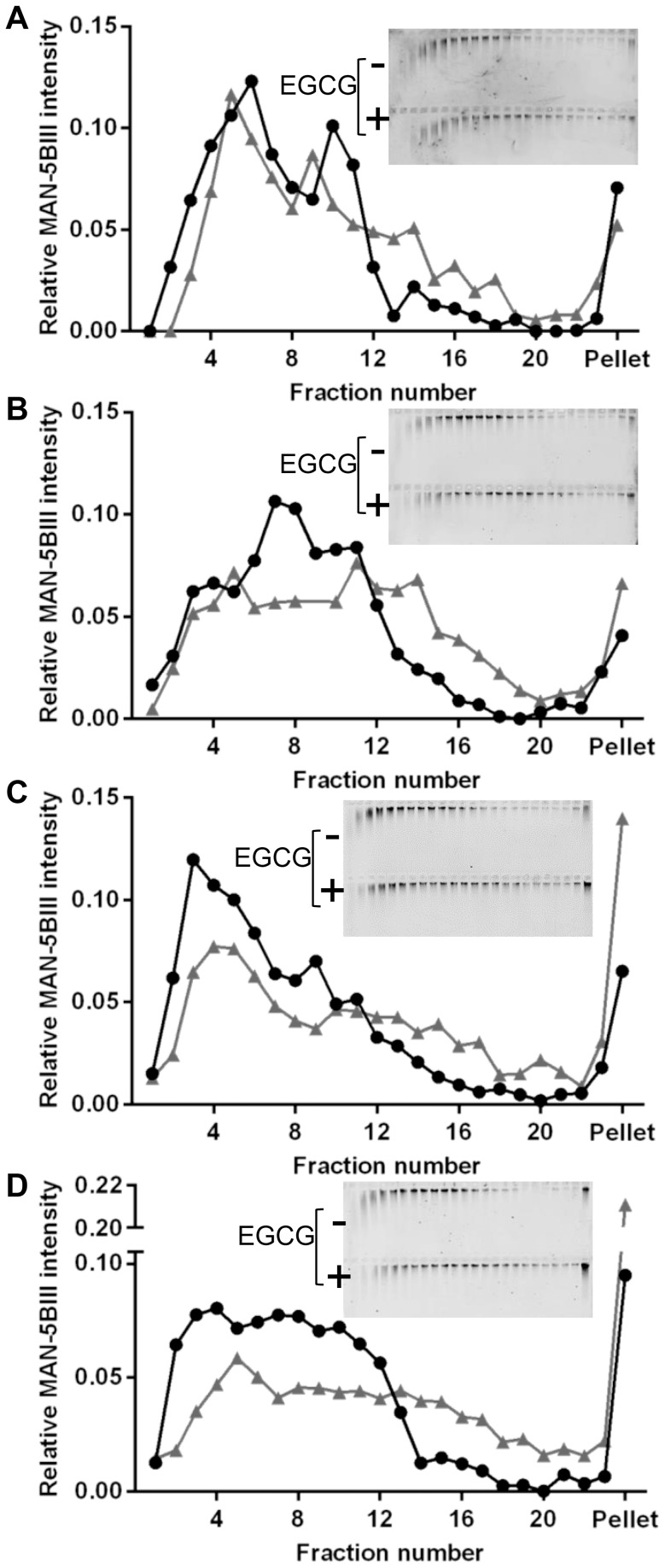
Figure 2. Increased sedimentation rate of purified MUC5B in the presence of 1 mM EGCG.
Rate-zonal centrifugation of MUC5B (A: 200 µg/mL, B: 400 µg/mL, C: 800 µg/mL, D: 1.6 mg/mL) with 1 mM EGCG (grey triangles) or PBS (black circles), performed in 10–35% (w/v) sucrose gradients in PBS. MUC5B was detected by agarose gel electrophoresis and western blotting with the MAN-5BIII antiserum (insets; MUC5B (top panel) and MUC5B+EGCG (bottom panel)). Bands were quantified using the Odyssey Imaging system.

