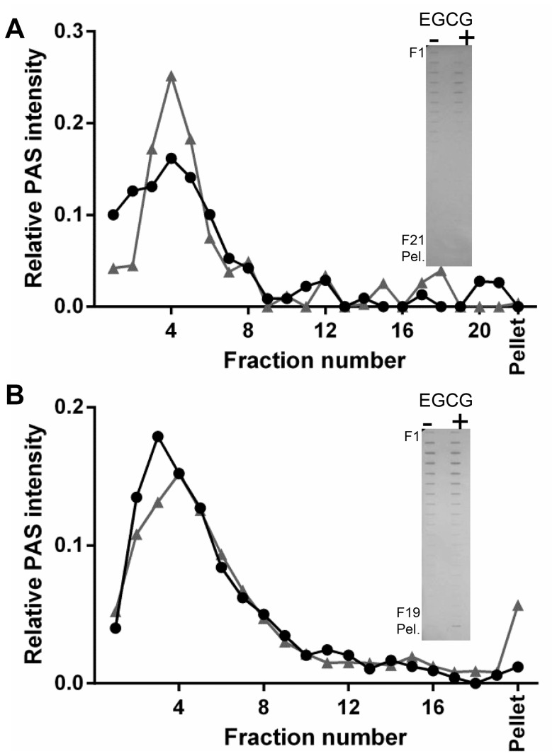
Figure 6. MUC5B oligosaccharide-rich regions do not aggregate in the presence of excess EGCG.
MUC5B T-domains were generated by trypsin-digestion and made to 200 µg/mL (A) and 600 µg/mL (B) before rate-zonal centrifugation with PBS (black circles) or 4 mM EGCG (grey triangles), in 5–20% (w/v) sucrose gradients. EGCG was removed from fractions using a HiTrap desalting column, fractions were slot blotted and PAS stained to detect glycoprotein (inset). Blots were scanned using Biorad ChemiDoc MP Imaging System and intensities measured using ImageLab software.

