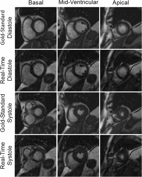
Figure 2.

Example images from a patient acquired using both the gold-standard breath-hold and ECG-gated cine (first and third rows) and the real-time imaging technique (second and fourth rows) for three different slice locations in both end-diastole and end-systole. Ejection fraction values of 68.8% and 66.7% were determined from the standard and real-time images, respectively. Both sets of images were rated to be artifact-free, and the gold-standard images were rated as “excellent” in all categories by both reviewers. The real-time images were rated as “excellent” in every category by Reviewer 1, and “excellent” in three categories and “good” in the remaining three by Reviewer 2.
