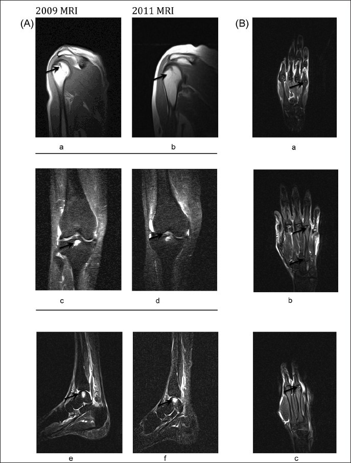
Fig.

(A). Persisting Focal Altered signal intensity lesions; (a, b) hypointense on T1 weighed sequence in posterior aspect of head of humerus; (c, d) lesion in intercondylar region of tibia; (e, f) Right Ankle. Fig. (B). (a) Lesion in 5th metacarpal with minimal effusion in MP joints of 2nd and 5th fingers at the end of 10th month of post CHIKV infection (2009). Lesions in sub-articular region of head of 3rd, 4th and 5th metacarpal with minimum effusion observed in 3rd to 5th MP joints, new erosion in 3rd and 4th metacarpals. (b, c) at the end of 3 years of post CHIKV infection (2011).
