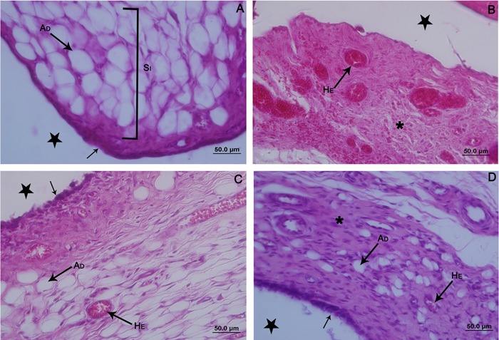
Figure 3. Photomicrographs of the synovial membrane of the ankle joint of Wistar rats. Sagittal section, hematoxylin and eosin staining. A: Control contralateral hindlimb. Membrane with thin synovial intima (black arrow), and subintima (SI) with predominance of fat cells (AD). B, G1 group, thickening of the synovial membrane, which was predominantly fibrous (asterisk) and with blood vessels (HE) full of red blood cells that spilled over the connective tissue. C, G2 group, synovial membrane with thickening in the apical region and intima with disorganized synoviocytes (black arrow). Presence of adipocytes (AD) in the subintima, with a moderate amount of red blood cells within the blood vessels (HE). D, G3 group, synovial membrane with areas of reorganization of the intima (black arrow). Subintima less fibrous (asterisk), with fat cells (AD) and few red blood cells in the blood vessels (HE). Articular cavity (star). G1: immobilized; G2: remobilized freely for 14 days; G3: remobilized by swimming and jumping in water for 14 days.

