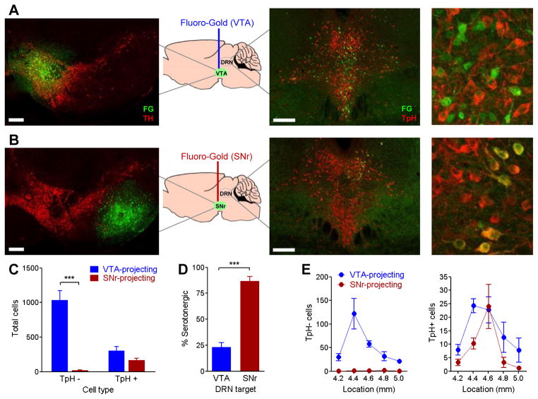
Figure 6. The majority of DRN cell bodies that project to VTA are non-serotonergic.
The retrograde tracer Fluoro-Gold was iontophoretically infused into the (A) VTA or (B) substantia nigra reticulata (n=4/group). Left panels, Fluoro-Gold (green) at infusion site, double-labeled with tyrosine hydroxylase (TH) to label dopamine neurons (red). Right panels, retrograde-labeled cells in DRN, double-labeled with tryptophan hydroxylase (TpH) to label serotonin neurons (red). Scale bars = 200 μm. C, Number of Fluoro-Gold-labeled cells in DRN tissue from mice injected with Fluoro-Gold in VTA or substantia nigra reticulata. Fluoro-Gold cells were grouped by presence or absence of tryptophan hydroxylase double-label (TpH+, TpH−). Two-way ANOVA (region x TpH label interaction) F(1,12)=34.11, p<0.0001; *** p<0.001 post-hoc. D, Percent of Fluoro-Gold labeled cells double-labeling for tryptophan hydroxylase. *** p<0.0001. E, Number of TpH− (left) and TpH+ (right) Fluoro-Gold labeled cells across the rostrocaudal axis of the DRN. X-axis indicates location of DRN tissue, in millimeters posterior to bregma.

