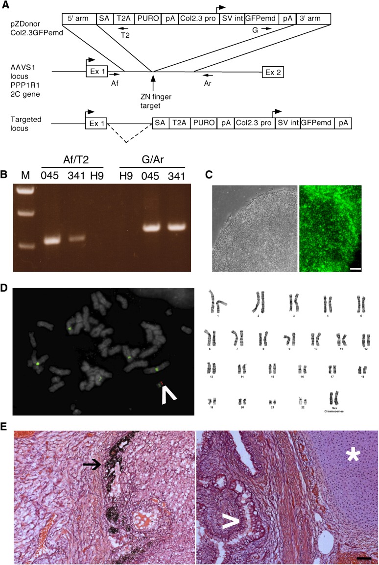
Figure 1.
Pluripotency of human embryonic stem cells (hESCs) is maintained after zinc finger nuclease targeting and single-cell cloning. (A): Strategy for targeting Col2.3GFPemd to the PPP1R12C gene at the AAVS1 locus. The vertical arrow shows the cut site of the zinc finger nuclease, where the transgene is inserted. The top diagram shows the targeting vector. The 5′ arm is the 5′ homology arm; T2A is the ribosome skip sequence from Thosea asigna, which separates the peptide sequence of the first exon of the PPP1R12C gene from PURO, the puromycin resistance gene. The box on the left labeled pA indicates bovine growth hormone polyadenylation signal, whereas the box on the right labeled pA indicates the SV40 virus polyadenylation signal. The targeted locus diagram shows that the puromycin resistance gene is driven by the PPP1R12C gene promoter, and the dashed line indicates splicing from the PPP1R12C gene first exon to the splice acceptor in the inserted construct. (B): AAVS1 forward and T2A reverse primer pairs showing the correct integration of the 5′ end of the construct; green fluorescent protein (GFP) forward and AAVS1 reverse primer pairs showing correct integration of the 3′ end of the construct. (C): A typical hESC colony from the C341-6 clone with a well-defined edge was imaged with phase contrast optics (left), and the C341-6 clone was stained with Alexa 445-labeled Tra1-60 antibody (right). Scale bar = 100 μm. (D): The single correct targeting site was demonstrated by fluorescence in situ hybridization on C341-6 clone with Alexa Fluor 555-labeled GFPemd probe (left; red band indicated by white arrowhead). The green signal is fluorescein-labeled Aquarius Enumeration Probe that hybridizes to the centromeres of chromosomes 1, 5, and 19. Light 4′,6-diamidino-2-phenylindole stain (gray) allowed visualization of chromosome banding pattern to confirm integration of transgene at 19q13.3–13.4. The C341-6 clone with normal karyotyping is demonstrated by Giemsa chromosome banding (right). (E): Tissues from three germ layers were found in a teratoma derived from C341-6 cells: melanocytes (ectoderm, left, black arrow), cartilage (mesoderm, right, white star), and intestine-like (endoderm, right, white arrowhead). Scale bar = 100 μm. Abbreviations: 045, colony C045; 341, colony C341; Af, AAVS1 forward primer pair; Ar, AAVS1 reverse primer pair; Col2.3 pro, 2.3-kilobase rat Col1a1 promoter fragment; Ex 1, exon 1 of the PPP1R12C gene; Ex 2, exon 2 of the PPP1R12C gene; G, GFP forward primer pair; GFPemd, GFPemerald; H9, H9Zn2.3GFP cells; pA, polyadenylation signal; SA, artificial splice acceptor; SV int, SV40 virus late intron; T2, T2A reverse primer pair; ZN, zinc.

