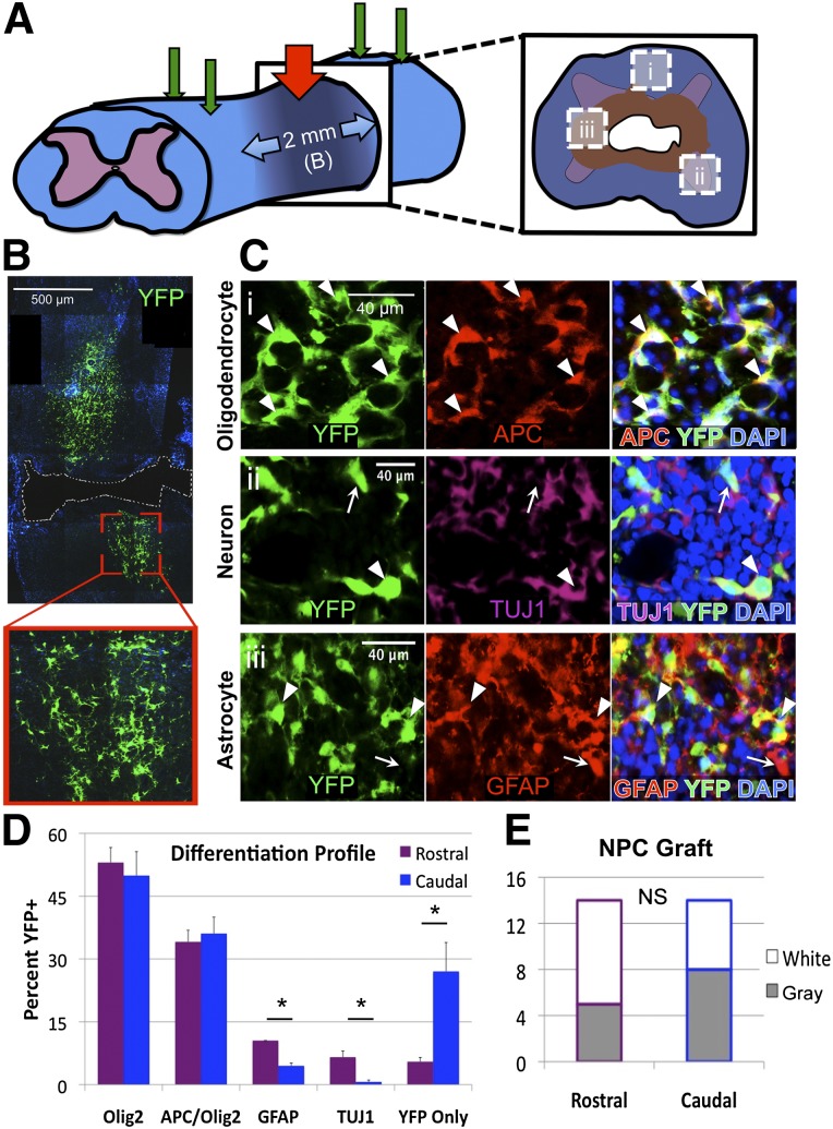
Figure 1.
Adult NPCs readily differentiate and engraft in the injured cervical spinal cord. (A): Schematic of the experimental design including 28-g cervical spinal cord injury at C6 (red arrow) and four NPC transplantation sites (green arrows). (B): Transplanted adult NPCs engraft within the injured cervical cord with long-term survival and cellular processes and morphology typical of NPC-derived progeny. (C): Differentiated NPCs and progeny (arrowheads) engraft and intersperse with endogenous cells (arrows) at 10 weeks after transplant. Transplanted cells differentiate to express markers of mature oligodendrocytes (Ci), neurons (Cii), and astrocytes (Ciii) (as shown in [A]). (D): Differentiation profiles of grafted cells at 10 weeks after transplant exhibit a majority of oligodendroglia (49.0%) and mature oligodendrocytes (34.6%), with neurons and astrocytes identified more in the rostral versus caudal region (6.6% vs. 0.7% neurons and 10.5% vs. 4.5% astrocytes). (E): The total YFP+ grafts observed on long-term follow-up indicate no difference between rostral and caudal regions within white matter (open) or gray matter (shaded). ∗, p < .05; NS, p > .05. Abbreviations: DAPI, 4′,6-diamidino-2-phenylindole; NPC, neural stem/precursor cell; NS, not significant; TUJ1, neuronal βIII-tubulin; YFP, yellow fluorescent protein.

