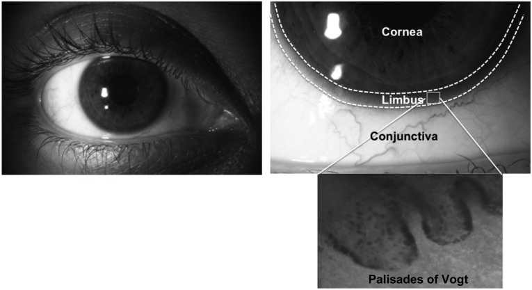
Figure 1.
Anatomical location of limbal stem cells. This figure shows the region of the limbus (marked with dashes lines) located between the avascular, transparent cornea and the vascularized, nontransparent conjunctiva. In this limbal region, the corneal stem cells are located within finger-like projections called the palisades of Vogt, as can be seen in the phase-contrast image.

