Figure 2.
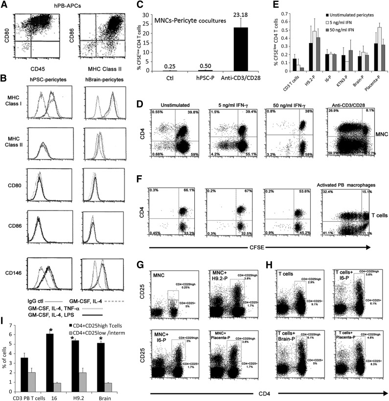
Allostimulation of peripheral blood mononuclear cells (PBMCs) or PB T cells with hPSC pericytes does not induce T-cell activation and increases the frequency of the CD4+CD25high T-cell subset. (A, B): Representative expression patterns of MHC class I, MHC class II, CD80, and CD86 by mature PB-derived macrophages (A) compared with hPSC and brain pericytes (B) under culture conditions (interleukin-4, granulocyte-macrophage colony-stimulating factor, and LPS/tumor necrosis factor-α) that gradually induce maturation of professional antigen presenting cells. (C–F): Proliferation of carboxyfluorescein succinimidyl ester-labeled CD4+ T cells after 72 hours in cocultures of PBMCs (C, D) or resting CD3+ PB T cells (E, F) with pericytes from the indicted sources. Activation of PBMCs with soluble antibodies against CD3/CD28 (C, D) and activation of PB T cells by allogeneic PB-derived APCs (F) served as positive controls. Data are mean ± SEM of three independent experiments and at least two different PB cells per experiment. (G, H): Representative fluorescence-activated cell sorting dot plots of expression of CD4 and CD25 on PBMCs (G) or PB T cells (H) following cocultivation with pericytes for 5 days. The frequencies of gated CD4+CD25+ and CD4+CD25high subsets are displayed. (I): Proportions of CD4+CD25+ or CD4+CD25high cell populations out of CD3+ PB T cells after 72 hours in pericyte cocultures compared with nonstimulated CD3+ PB T cells. Data are mean ± SEM of three independent experiments per pericytic line. ∗, p < .0001 compared with unstimulated T cells of the matched subset. Abbreviations: APC, antigen-presenting cell; Ctl, control; hPB, human peripheral blood; hPSC, human pluripotent stem cell; IFN, interferon; LPS, lipopolysaccharide; MHC, major histocompatibility complex; MNC, mononuclear cell; P, pericyte; PB, peripheral blood.

