Figure 3.
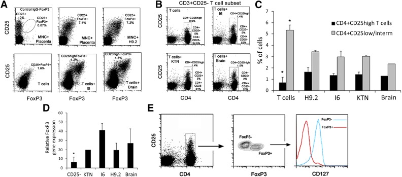
Pericytes from hPSC, brain, and placenta promote the formation of CD4+CD25highFoxP3+CD127− T cells. (A): Representative dot plots of CD25 and FoxP3 expression of gated CD4 T cells from cocultures of peripheral blood mononuclear cells or peripheral blood (PB) CD3+ T cells with hPSC, brain, and placenta pericytes after 3 days of induction. (B): CD3+ T cells were separated into CD3+CD25+ and CD3+CD25− subsets, and then the CD3+CD25− subset was used for pericyte induction experiments. The expression of CD4 and CD25 and the formation of CD4+CD25high populations among the allostimulated CD25− T-cell subset are shown in representative dot plots. (C): Percentages of CD4+CD25+ and CD4+CD25high cell populations generated from the CD3+CD25− T-cell subset in hPSC and brain pericyte cocultures. Mean ± SEM of triplicate samples is shown from seven independent experiments with seven different PB CD3+CD25− T cells and at least four pericyte cell lines per experiment. ∗, p < .05 compared with pericyte-stimulated T cells of a matched subset. (D): Quantitative polymerase chain reaction gene expression of FoxP3 in the CD3+CD25− T-cell subset compared with the CD3+CD25− T-cell subset after 3 days of cultivation with hPSC and brain pericytes. Data are mean ± SEM of 3 independent experiments with PB cells from 2 different donors. ∗, p = .05 compared with pericyte-stimulated CD25− T cells. (E): Representative expression of FoxP3 in gated CD4+CD25+ population, which was induced by pericyte from the CD3+CD25− subset within 72 hours. CD127 expression was evaluated on CD4+CD25+FoxP3+ and CD4+CD25+FoxP3− subpopulations. Abbreviation: MNC, mononuclear cell.

