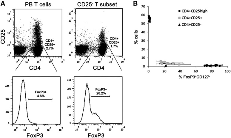
Figure 6.
Positive correlation between pericyte allostimulated expression of CD25 and FoxP3 in converted CD4 T cells. (A): Representative flow cytometry analyses of the expression of FoxP3 in gated CD4+CD25+ subsets that were derived from either PB CD3 T cells (left panels) or isolated CD4+CD25− subset (right panels), which was allostimulated for 72 hours with pericytes. (B): CD25 and FoxP3 expression-based clustering of pericyte-stimulated CD4+CD25− T cells. The nonclustered CD4+CD25high black dot represents T cells that were treated with transforming growth factor-β RK inhibitor in the absence of pericytes. n = 3 lines of hPSC, brain, and placenta pericytes with duplicates and 5 different donors. Abbreviation: PB, peripheral blood.

