
Figure 4.
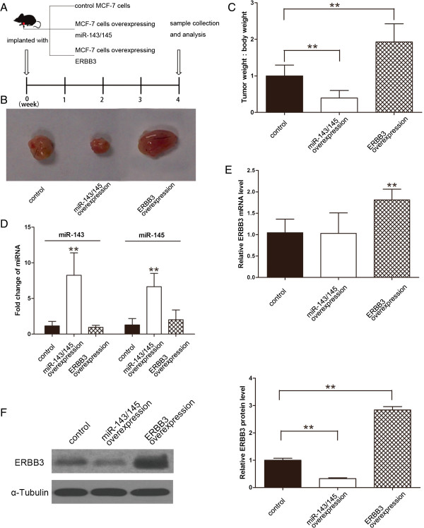
Effects of overexpression of miR-143/145 or ERBB3 on the growth of breast cancer cell xenografts in mice. (A) Flow chart of the experimental design. MCF-7 cells were either infected with a lentiviral expression vector to express miR-143 and miR-145 or transfected with an ERBB3 plasmid to overexpress ERBB3. MCF-7 cells (1 × 10⁷ cells per 0.1 mL) with increased miR-143/145 or ERBB3 levels were then implanted subcutaneously into 4-week-old C57/BL6 mice (7 mice per group), and tumor growth was evaluated at day 28 after cell implantation. (B) Representative image of the tumors from mice implanted with the control MCF-7 cells, the miR-143/145-overexpressing MCF-7 cells or the ERBB3-overexpressing MCF-7 cells. (C) Quantitative analysis of the tumor weights from mice implanted with control MCF-7 cells, miR-143/145-overexpressing MCF-7 cells or ERBB3-overexpressing MCF-7 cells. (D) Quantitative RT-PCR analysis of miR-143 and miR-145 levels in the tumors from the mice implanted with control MCF-7 cells, miR-143/145-overexpressing MCF-7 cells or ERBB3-overexpressing MCF-7 cells. (E) Quantitative RT-PCR analysis of ERBB3 mRNA levels in the tumors from mice implanted with control MCF-7 cells, miR-143/145-overexpressing MCF-7 cells or ERBB3-overexpressing MCF-7 cells. (F) Western blotting analysis of ERBB3 protein levels in the tumors from mice implanted with control MCF-7 cells, miR-143/145-overexpressing MCF-7 cells or ERBB3-overexpressing MCF-7 cells. Left panel: representative image; right panel: quantitative analysis. * P < 0.05; ** P < 0.01.