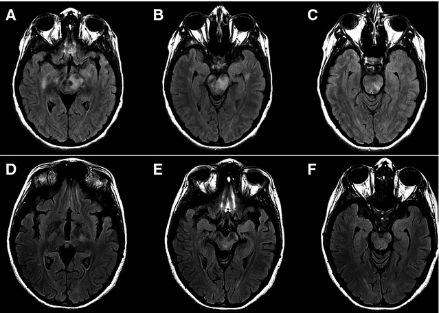
Figure 1.

(A–C) MRI on axial Fast-FLAIR sequence shows demyelinating areas involving gyri rectus, mesencephalon, and pons. (D–F). Control MRI performed 15 months after initial symptoms shows improvement in demyelinating lesions and secondary cortical atrophy.
