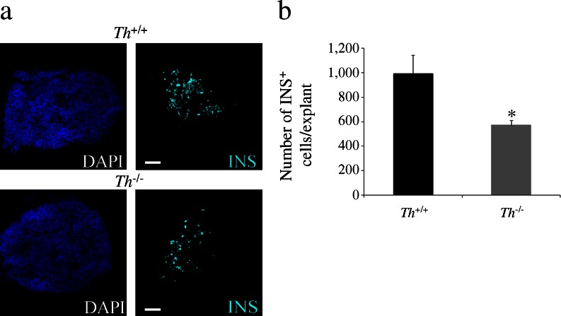
Fig. 3.
Decreased number of insulin-expressing cells in the TH-deficient pancreatic explants. (a) Immunostaining for insulin (INS, cyan) in E13.5 pancreatic explants cultured for 5 days. Nuclei are stained with DAPI. Scale bar, 100 μm. (b) Quantification of total number of insulin-expressing cells. Results represent the mean ± SEM of at least four explants per genotype. *p < 0.05 vs Th +/+

