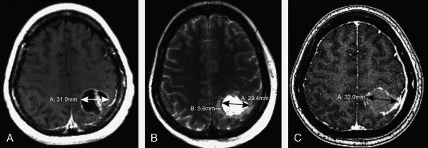
Figure 3. 41-year-old female with breast cancer metastatic to the brain A.

Axial post-contrast T1-weighted image shows a 31-mm enhancing, partially cystic, left parietal mass. B. Axial T2WI from the post-operative MRI obtained within 24 hours shows a 29-mm resection cavity. Minimal surrounding edema measures 6 mm. C. Axial post-contrast T1WI obtained 18 days post-surgery shows the resection cavity diameter increased to 32 mm.
