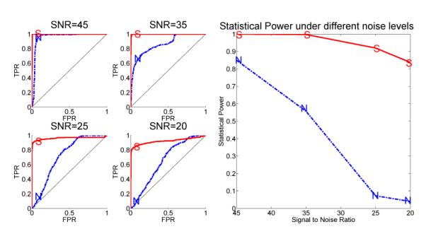
Figure 5.

The advantage in ROC curves and statistical power of the SC (marked by “S”) versus the NC methods (marked by “N”) to detect differential interactions in the cdc2-cyclin cell division cycle model. Left: The ROC curves under different noise levels. Right: The statistical power of both methods as a function of the noise level.
