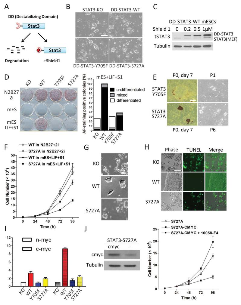
Figure 1.
Diverse functions of STAT3 at different phosphorylation sites revealed using transgenic STAT3 in STAT3−/− mouse embryonic stem cells (mESCs). (A): Schematic diagram showing the principle of destabilizing domain (DD)-STAT3 expression system. (B): STAT3−/− + DD-STAT3 transgenic mESCs in N2B27 + 2i medium display typical mESC morphology. (C): Dose-dependent modulation of DD-STAT3 protein level by Shield 1 (S1). Cells were cultured in N2B27 + 2i medium and treated with various concentrations of S1 overnight. DD-STAT3-WT expression in STAT3−/− mESCs is depicted as an example. The two bands in the STAT3 blot represent STAT3 from mouse embryonic fibroblast feeder cells (lower band) and DD-STAT3 fusion protein (upper band). (D): Comparison of self-renewal potential of different STAT3−/− + DD-STAT3 cells under various culture conditions: N2B27 + 2i, mESC, and mESC+LIF+S1. We counted the numbers of differentiated, undifferentiated and mixed colonies these mESCs formed after 7 days in the mESC+LIF+S1 culture condition. (E): Alkaline phosphatase staining and phase contrast images showing the short and long-term self-renewal potential of STAT3−/− + DD-STAT3-Y705F and STAT3−/− + DD-STAT3-S727A mESCs under the mESC+LIF+S1 condition. (F): Growth curves of STAT3−/− + DD-STAT3-WT and STAT3−/− + DD-STAT3-S727A mESCs in mESC+LIF+S1. Their growth curves in N2B27 + 2i are used as positive controls. (G): Representative images showing different STAT3−/− + DD-STAT3 mESCs plated at a density of 1 × 10⁴ cells per square centimeter on gelatin-coated plates and cultured in mESC+LIF+S1 overnight. (H): TUNEL-FITC staining of STAT3−/− + DD-STAT3 mESCs in mESC+LIF+S1. (I): Myc transcription level in STAT3−/− + DD-STAT3 mESCs in mESC+S1 after 1 hour of leukemia inhibitory factor (LIF) stimulation. (J): Improved proliferation of STAT3−/− + DD-STAT3-S727A mESCs with forced expression of c-myc: left: western blot showing overexpression of c-myc; right: growth curve of cells in mESC+LIF+S1 condition, 10058-F4: c-myc inhibitor (50 μM) (B, E, G, H) Scale bars = 50 μm. Abbreviations: DD, destabilizing domain; KO, knock out; LIF, leukemia inhibitory factor; mESC, mouse embryonic stem cell; WT, wild type.

