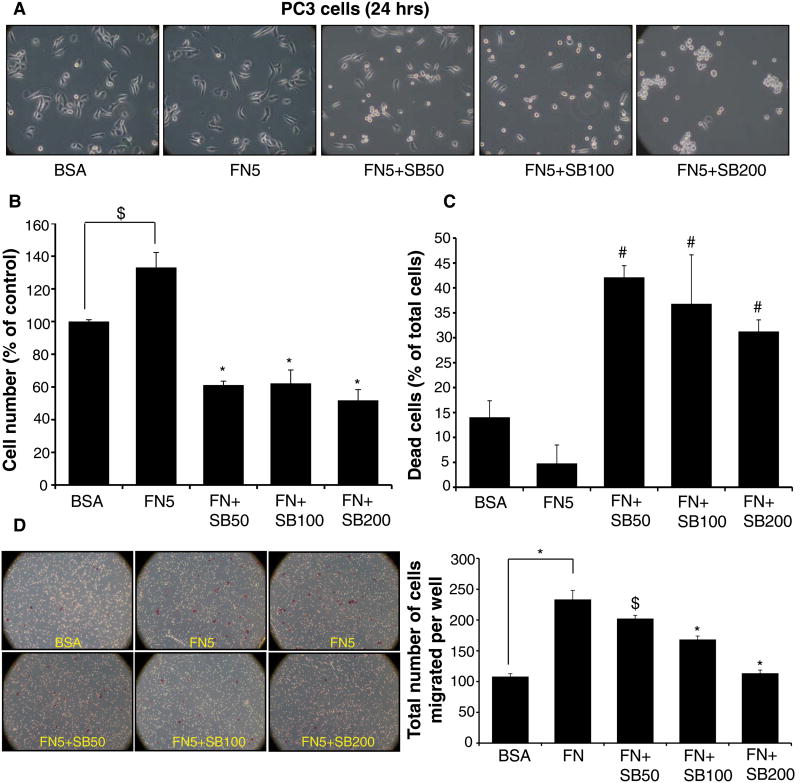
Figure 2. Silibinin inhibits fibronectin-induced proliferation and motility in PC3 cells.
PC3 cells were cultured in RPMI media (with 0.5% FBS) for 24 hrs; thereafter cells were collected and re-plated on fibronectin (5 μg/ml) coated plates with or without silibinin (50-200 μM). PC3 cells plated on BSA (5 μg/ml) coated plates served as relevant control. After 24 hrs, cells were analyzed for (A) morphology under a microscope; and (B-C) total cell number and dead cells (% of total cells). (D) Effect of silibinin treatment on fibronectin-induced motility was analyzed in a Transwell migration assay. Data shown in bar diagram is mean±SEM of three samples.
Abbreviations: BSA: Bovine serum albumin; FN: Fibronectin; SB: Silibinin. *, p≤0.001; #, p≤0.01; $, p≤0.05

