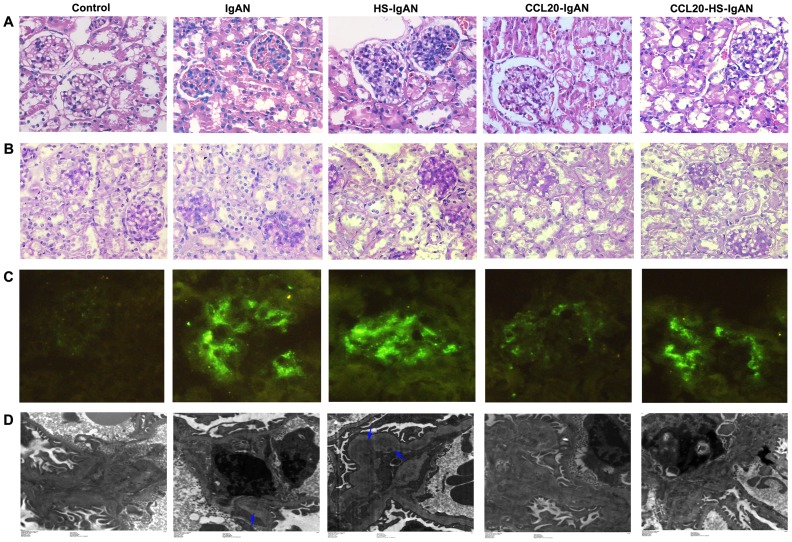
Figure 2. HS worsens and CCL20 antibody improves renal damage in IgAN mice.
Representative images of HE-stained (A, 400×), PAS-stained (B, 400×), Immunofluorescence (C, 200×) and transmission electron micrographs (D) kidney sections from mice as indicated. For immunofluorescence staining, IgA antibody was used. The arrows in D point to high electron dense deposition in glomerular mesangial region.

