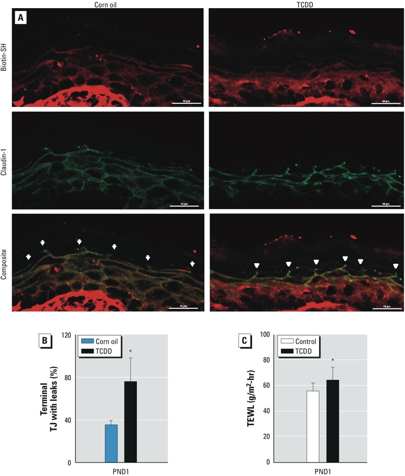
Figure 4.

Disruption of TJ permeability barrier after in utero TCDD exposure. (A) Photomicrograph of PND1 murine skin exposed to corn oil or TCDD. Arrows indicate claudin-1–positive sites with biotin-SH stops, and arrowheads indicate claudin-1–positive sites without biotin-SH stop. Bars = 10 μm. (B) Quantification of claudin-positive sites for terminal TJs without biotin-SH stops (≥ 3 visual fields per sample were counted, and 3 pups per treatment condition were analyzed). A total of 87 terminal TJs were counted in corn-oil samples; 30 of these terminal TJs had leaks for biotin-SH. In TCDD-exposed pups, 104 terminal TJs were counted, and 72 of these terminal TJs had leaks for biotin-SH. (C) TEWL after SC removal in PND1 mice. The dorsal skin of mice was tape stripped six times to remove most of the SC before TEWL was measured. At least 22 pups from 4 dams were assayed in the control or TCDD-exposed group. Values are means ± SDs. *p < 0.05, compared with age-matched control samples by Student’s t-test.
