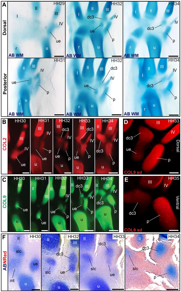
Figure 6. Loss of the ulnare and late formation of distal carpal 3 (“element x”) in the chicken.
(A) Whole-mount alcian blue staining confirms the ulnare is the first carpal formed in avian embryos, distal to the ulna. Thereafter, a distal carpal 3 (referred to as “element x” in previous embryological descriptions) is formed distal to the ulnare, coexisting with it. Finally, the ulnare disappears, whereas dc3 persists. (B) Collagen type II and (C) collagen type IX whole-mount immunostaining documents the formation of dc3 distal to the ulnare and the reduction and disappearance of the ulnare. (D) Detail of dc3 and receding ulnare, coexisting in the chicken embryo, as observed by spin-disc microscopy. See Video S2. (E) Detail of dc3 after disappearance of the ulnare. The dc3 cartilage thereafter acquires a bent, “v”-like shape in galloanserae (chicken and duck), but not other bird species observed (Video S3). (F) Histological sections showing the late formation of dc3, its co-existence with the receding ulnare, and the disappearance of the ulnare in the chicken embryo. Scale bars, (A–C and F) 300 µm and (D and E) 150 µm.

