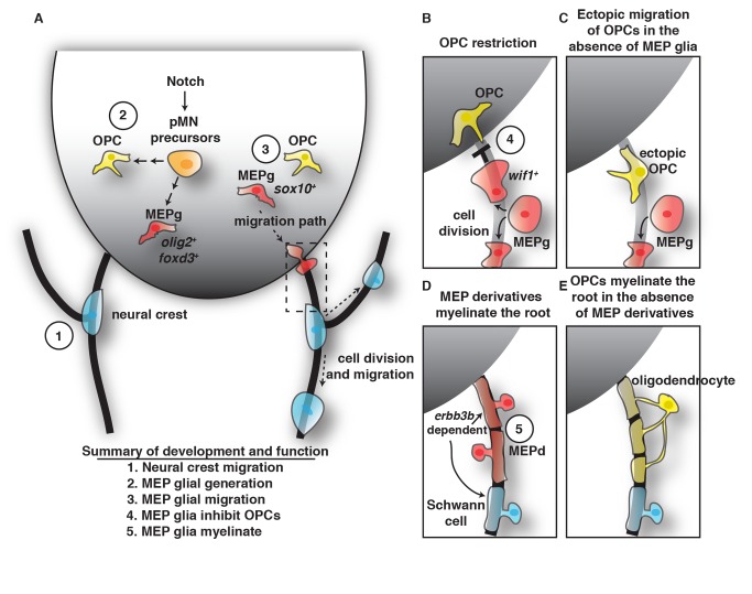
Figure 10. Summary of MEP glial development and function.
(A) Schematic of a cross-section of the spinal cord summarizing the findings that pMN precursors (orange cells) generate OPCs (yellow cells) and olig2/foxd3 expressing MEP glia (MEPg) (red cells). These precursors are dependent on Notch signaling. MEPg then initiate sox10 expression, migrate to the MEP (dashed box), squeeze through the TZ, and reside along spinal motor root axons. (B–D) Inset of the MEP depicted as dashed box in (A). (B) OPCs are restricted from entering the PNS by wif1+ MEPg. (C) In the absence of MEPg, OPCs ectopically migrate out of the spinal cord. (D) MEP glial derivatives (MEPd) myelinate the root and in their absence, and (E) oligodendrocytes myelinate motor root axons with central myelin. The association of MEPd and Schwann cells with the root is erbb3b dependent. Each developmental stage of MEPg is numerically labeled, showing MEPg (1) are independent of neural crest, (2) are generated from ventral spinal cord precursors, (3) migrate into the periphery via the MEP TZ, (4) inhibit OPC migration, and (5) myelinate the root.

