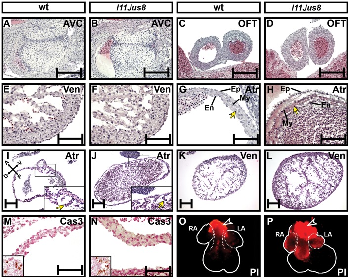
Figure 2. Effect of the homozygous Erbb2M802R mutation on heart morphology in l11Jus8 hearts.
(A–L) Representative H and E stained coronal sections of E12.5 hearts. (A–B) Atrioventricular cushions, (C–D) dorsal outflow tract, (E–F) ventricles, (G–H) atria. Arrows in (G–H) point to the atrial wall. AVC, atrioventricular cushion; OFT, outflow tract; Ven, ventricle; Atr, atrium; Ep, epicardium; En, endocardium; My, myocardium. (I–J) Longitudial atrial and (K–L) ventricular sections; homozygous l11Jus8 heart had distended atria. Embryos in I–L are littermates. Magnified areas in (I–J) show the developing pectinate muscles (arrows). (M–N) Activated Caspase 3 (Cas3) staining of atrial wall at E11.5. Inset shows positive activated Caspase3 staining from trigeminal ganglion on same embryo section. (O–P) Propidium Iodide (PI) labelling of the necrotic cells in E12.5 hearts. RA, right atrium; LA, left atrium. Arrowheads point to the unspecific PI staining resulting from cells of the OFT damaged during dissection. Scale bars: 200 µm in A–H and M–N, 50 µm in I–L.

