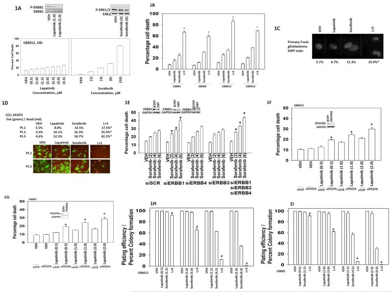
Figure 1. Sorafenib and lapatinib interact to kill glioblastoma cells.
(A) GBM12 cells were treated with sorafenib (1.0-10.0 μM) or lapatinib (0.5-3.5 μM) as indicated. Cells were isolated 24h after exposure and viability determined by trypan blue exclusion (n = 3, +/− SEM). Upper blots: lapatinib reduces ERBB1 phosphorylation; sorafenib modestly reduces ERK1/2 phosphorylation. (B) GBM5 / GBM6 / GBM12 / GBM14 cells were treated with vehicle (DMSO), sorafenib (6.0 μM) and/or lapatinib (2.0 μM) as indicated. Cells were isolated 24h after exposure and viability determined by trypan blue exclusion (n = 3, +/− SEM) *p < 0.05 greater than vehicle control. (C) Freshly isolated glioblastoma cells from the operating room were treated with vehicle (DMSO), sorafenib (3.0 μM) and/or lapatinib (2.0 μM) as indicated. Cells were isolated 24h after exposure and spun onto glass slides and DAPI stained. Representative images are shown from each treatment condition. (D) Low passage primary human glioblastoma cells (patient 1; patient 2; patient 3) were treated with vehicle (DMSO), sorafenib (3.0 μM) and/or lapatinib (2.0 μM) as indicated. Cells were isolated 24h after exposure and viability determined by live = green / dead = red assay using a Hermes WiScan platform (n = 3, +/− SEM) *p < 0.05 greater than vehicle control. (E) GBM12 cells were transfected to knock down the expression of ERBB1, ERBB3 and/or ERBB4. Thirty six h after transfection cells were treated with vehicle (DMSO), or sorafenib (2.0-6.0 μM) as indicated. Cells were isolated 24h after exposure and viability determined by trypan blue exclusion (n = 3, +/− SEM) *p < 0.05 greater than corresponding siSCR control. (F) GBM12 cells were transfected to knock down expression of PDGFRβ. Thirty six h after transfection cells were treated with vehicle (DMSO), or lapatinib (0.5-2.0 μM) as indicated. Cells were isolated 24h after exposure and viability determined by trypan blue exclusion (n = 3, +/− SEM) *p < 0.05 greater than corresponding siSCR control. (G) GBM5 cells were transfected to knock down expression of PDGFRα. Thirty six h after transfection cells were treated with vehicle (DMSO), or lapatinib (0.5-2.0 μM) as indicated. Cells were isolated 24h after exposure and viability determined by trypan blue exclusion (n = 3, +/− SEM) *p < 0.05 greater than corresponding siSCR control. (H) and (I) GBM12 or GBM5 cells, as indicated in the Figure, were plated (250-1,500 single cells / well) in six well plates. Cells were permitted to attach and after 12h treated with drugs. Cells were treated with vehicle (DMSO), sorafenib (1.5-6.0 μM) and/or lapatinib (0.5-2.0 μM) as indicated for 24h. Media was removed and replaced with drug free media and cells permitted to grow and form colonies for the next 14 days (n = 3 in sextuplicate, +/− SEM) #p < 0.05 less than vehicle control.

