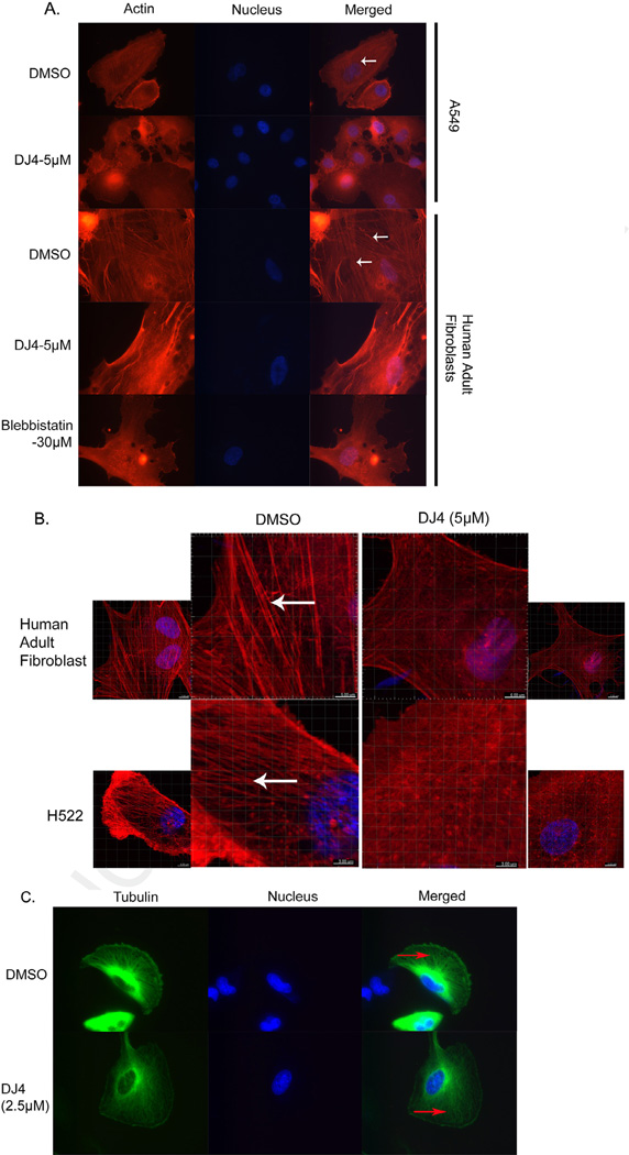
Fig. 4.
Cytoskeletal changes induced by DJ4 in A549, H522, U251 and human adult fibroblast cells after DJ4. (A) A549 and fibroblasts were treated with DJ4 for 1 h and stained with rhodamine-phalloidin and DAPI for visualization of stress fibers (red; indicated by white arrow) and nuclei (blue) respectively. Images were captured at 600× magnification using an inverted fluorescent microscope with an oil objective. (B) Normal human adult fibroblasts and H522 cells were treated with 5 µM DJ4 for 8 h and 3.5 h respectively. Cells were stained for stress fibers (red colored indicated by white arrow) with rhodamine-phalloidin and for nuclei (blue) with DAPI. Images were captured at 400× under a confocal microscope. (C) U251 glioblastoma cells stably expressing EGFP-Tubulin were treated with DMSO or DJ-4 (2.5 µM) for 4 h, fixed and nuclei were stained with DAPI. Red arrows indicate microtubules (green). Images were captured at 600× using an inverted fluorescent microscope with an oil objective. (For interpretation of the references to color in this figure legend, the reader is referred to the web version of this article.)

