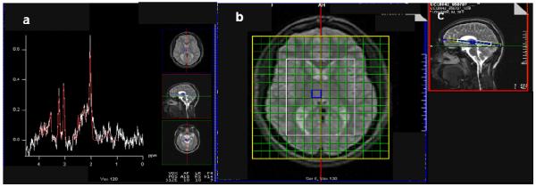
Fig. 1.

1H MRSI spectrum (a) acquired from right mesial thalamus of subject (PRESS, 1.5 T, repetition time/echo-time = 1500/30 ms). Glutamate and overlapping glutamine (together “Glx”) present as the shoulder in the 2.3-2.4 ppm range. Axial-oblique (b) and sagittal (c) MRI of the brain depicting 9-mm thick MRSI slab of 11×11-mm2 voxels. The PRESS volume (white; ~8×10 voxels) was aligned parallel to the genu-splenium line and fit to the subject. It sampled bilateral thalamus, and environs. The blue square in (b-c) denotes voxel yielding MR spectrum in (a).
