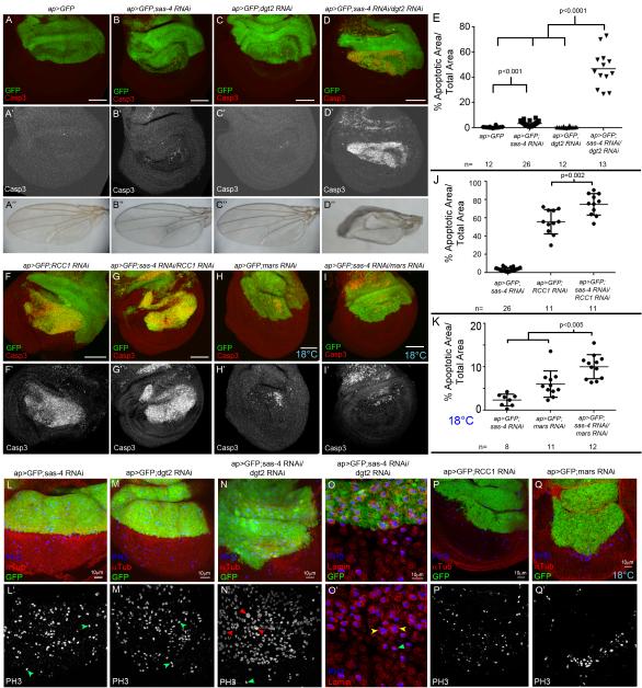
Fig3. Acentrosomal cells depend on alternative microtubule nucleation pathways.
(A-E, F-K). Apoptosis (Casp3), quantified in (E,J,K), (B“-D”) Adult wings. (A) Control=ap-Gal4 driving UAS-GFP (ap>GFP) in the dorsal compartment. (B) ap>GFP driving sas-4 RNAi. (C) ap>GFP driving Augmin component dgt2 RNAi. (D) Double Sas-4/Dgt2 knockdown. (F) Knockdown of RCC1 alone. (G) RCC1/Sas-4 codepletion (H) Knockdown of Mars at 18°C. (I) Mars/Sas-4 double knockdown at 18°C. (L,M) Sas-4 or Dgt2 knockdown in the dorsal wing. No change in the distribution of prophase, metaphase (green arrows), anaphase, or telophase cells. (N) Codepleting Sas-4 and Dgt2. Obvious enrichment of mitotic cells with virtually all cells stalled in prophase/prometaphase; note apparently polyploid nuclei (red arrows versus green arrow). (O) Sas-4/Dgt2 codepleted disc. Lamin staining (nuclear envelope) confirms that large mitotic nuclei are within a single cell (yellow versus green arrows). (P,Q) RCC1 or Mars knockdown. Fewer mitotic cells in knockdown region. See also Fig S3-S5.

