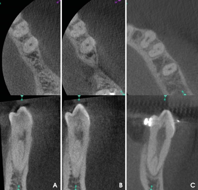
Fig. 2.
Axial (up) and buccolingual (down) CBCT images show the Vertucci type II configuration. B. Shapes not included in Vertucci's classification. One canal in the coronal third of the root, three canals in the middle third, and one canal in the apical third. C. Axial (up) and buccolingual (down) CBCT images show the Vertucci type V configuration.

