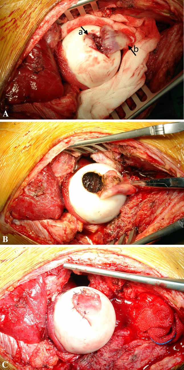
Fig. 1A–C.

(A) The femoral head is exposed after dislocation. Arrow a indicates where the articular cartilage is breached, which can help locate the tumor but makes coverage with the affected cartilage (trapdoor) impossible. Arrow b indicates the acetabulum end of the ligamentum teres. (B) One window is open near the ligamentum teres, and the entire tumor can be curetted via this window. (C) After bone grafting, the ligamentum teres is sutured together with the cartilage rim of the window cartilage.
