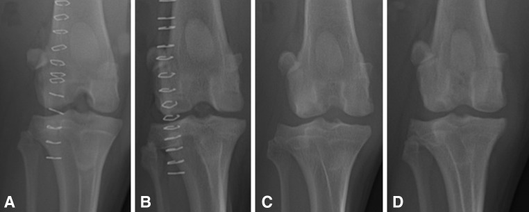
Fig. 3A–D.

The radiographic appearances of the knees of (A) Dog 1 and (B) Dog 5 immediately after implantation of osteochondral allografts are shown. These radiographs show the appearances of the knees of (C) Dog 1 and (D) Dog 5 6 months after implantation of osteochondral allografts into the medial and lateral femoral condyles. For both of these dogs, MOPS-60 grafts were placed in the medial femoral condyles and SOC-60 grafts were placed in the lateral femoral condyles. Osseous integration of SOC and MOPS grafts is apparent based on the loss of radiolucency at the graft margins. There was no radiographic evidence for failure of osseous integration for any graft.
