Fig. 1A–B.

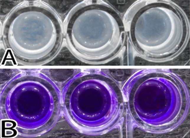
Images illustrate S epidermidis growth after 24 hours of exposure. (A) The biofilms present in the wells after removal of the medium are shown. (B) The resultant stain with crystal violet is shown.

Images illustrate S epidermidis growth after 24 hours of exposure. (A) The biofilms present in the wells after removal of the medium are shown. (B) The resultant stain with crystal violet is shown.