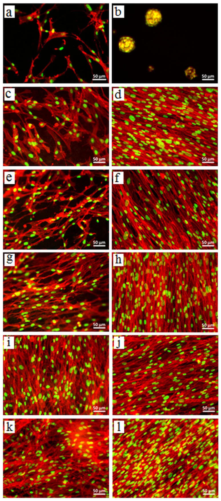
Figure 8. Morphological assessment of NHDF on hydrogels by analyzing F-actin pattern.

FM images of NHDF adhered on (a,b) alginate, (c,d) ADA70-GEL30, (e,f) ADA60-GEL40, (g,h) ADA50-GEL50, (i,j) ADA40-GEL60, and (k,l) ADA30-GEL70 after 4 days (left column) and 7 days (right column) of incubation. The cells were stained for F-actin (red) and nuclei (green). Scale bar: 50 µm.
