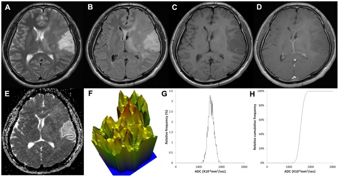
Figure 2. Images of a 43-year-old male with a grade II astrocytoma.
(A) T2-weighted image, (B) T2 FLAIR image, (C) T1-weighted image, (D) contrast-enhanced T1-weighted image, (E) ADC map with ROI placement, with the corresponding (E) 3-D height map of the ADC signal intensity, (G) histogram of ADC and (F) cumulative ADC histogram. The entropy value of ADC was 6.168.

