
Figure 7. Photobleaching of the retinal chroophore in cR3 and bR at 20°C.
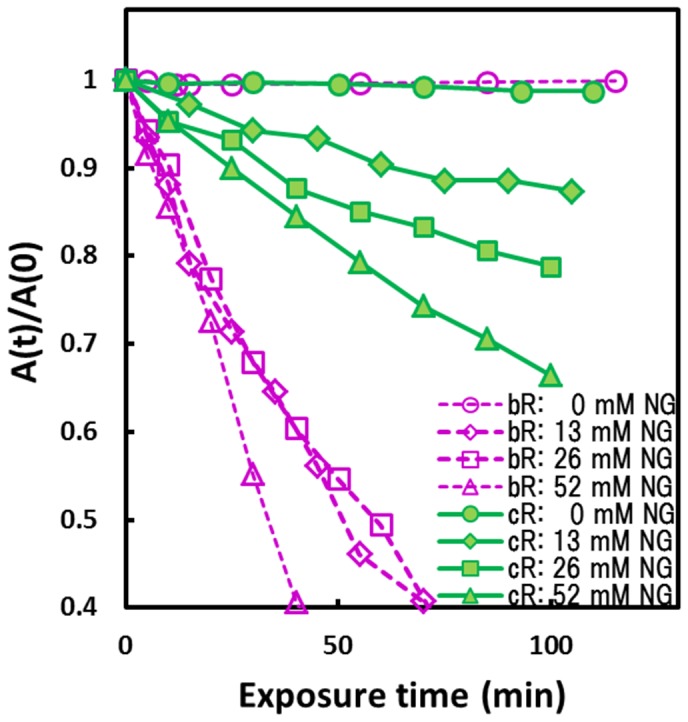
The solubilized sample was prepared by incubating cR3-rich claret membrane or the purple membrane of H. salinarum in a solution containing 0.01 M HEPES (pH 7) and 13–52 mM nonylglucoside (cmc ∼6 mM) at 30°C for 2–24 hours); unsolubilized membranes were removed by centrifugation at 100,000 rpm. The membrane suspension or the solubilized sample was exposed to strong orange light (570–700 nm, 80 mW/cm2). Absorption changes at 580 nm are plotted against the exposure time. Here, A(0) is the absorbance (∼0.35) observed just after the light-adaptation (i.e., after the pre-illumination for ∼5 minutes).