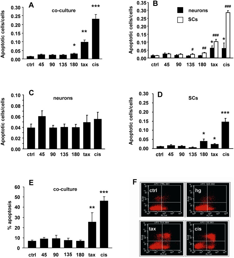
Figure 2. Hyperglycaemia did not increase DRG neuron or Schwann cell (SC) apoptosis.
(A) Neuron-SC co-cultures showed a modest increase of apoptosis rate only at the highest glucose concentration; paclitaxel (tax) and cisplatin (cis) exposition was used as positive controls. (B) In DRG neuron monocultures, hyperglycaemia did not induce apoptosis. (C) Tubulin-III and GFAP staining demonstrated that apoptosis mainly involved SC. (D) SC monocultures showed a mild increase of apoptosis rate at the highest glucose concentrations, similar to that observed in co-cultures (see C). (E) Flow cytometry by annexin V/PI assay confirmed the absence of apoptosis in co-cultures exposed to hyperglycaemia. (F) Representative cytogram showing the absence of apoptosis in control co-culture (ctrl) and after 24 hour exposition to hyperglycaemia 45 mM (hg), compared to the high apoptosis rate after exposition to anti-neoplastic compounds (tax, cis). Data are expressed as mean±SEM of independent experiments (n = 8) *p<0.05; **p<0.005; ***p<0.0005 vs controls; #p<0.05; ##p<0.005; ###p<0.0005 vs SC controls.

