
Figure 3. Reduced number of Th2 cells in the thoracic lymph nodes of PLCε ΔX/ΔX mice.
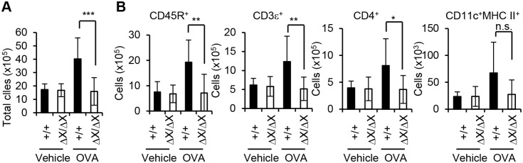
(A) Total leukocyte count of the thoracic lymph nodes. All visible thoracic lymph nodes were collected from the sensitized mice with the indicated PLCε genotype 1 day after the last challenge with vehicle alone or OVA (6 to 15 mice of each group), and subjected to isolation of leukocytes. Leukocyte number was determined using a hematocytometer. Data are expressed as the mean ± SD. ***, p<0.001. (B) Flowcytometric analysis of leukocytes. Collected leukocytes in (A) were further analyzed flowcytometrically for the expression of the indicated cell surface antigens. Data are expressed as the mean ± SD. *, p<0.05; **, p<0.01. n.s., statistically not significant.