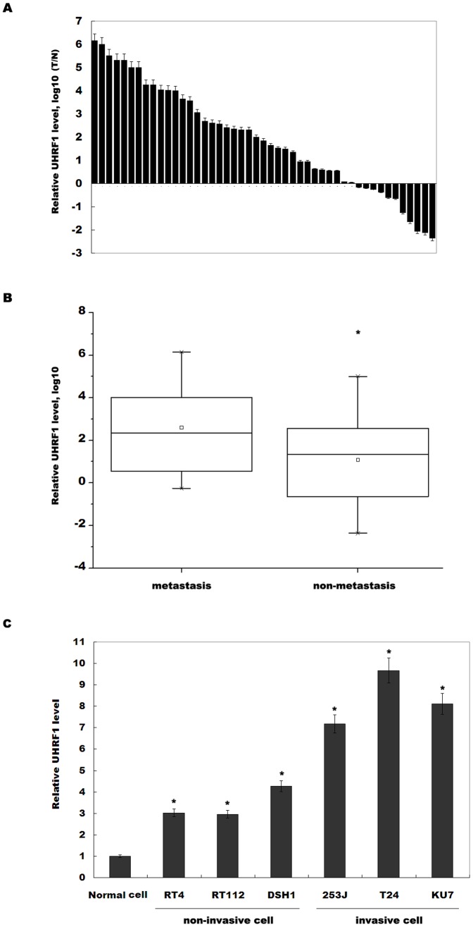
Figure 1. Higher UHRF1 expression is associated with bladder cancer metastasis.
(A) The quantitative analysis of UHRF1 expression level was carried out in bladder cancer tissues (n = 47) and adjacent normal tissues. Total RNA was extracted and subjected to real-time PCR to analyze the expression of UHRF1 in each sample. β-actin was used as an internal control. The relative UHRF1 level was calculated by 2−ΔΔCt where ΔCt = Ct (UHRF1) – Ct (β-actin) and ΔΔCt = ΔCt (tumor tissue) – ΔCt (adjacent normal tissue). (B) The bladder cancer specimens were divided into two groups based on clinical progression. The UHRF1 levels in the metastasis group (n = 25) were higher than those in the no-metastasis group (n = 22). *p<0.05. (C) UHRF1 expression level was assayed by real-time PCR in three noninvasive and three invasive bladder cancer cell lines. Normal urothelial cells were used as control. *p<0.05.

